
Vote for the 2025 People’s Choice Award
Voting Closes November 2nd – The following list is alphabetically organized by researcher’s last name.
Cochlear Hair Cells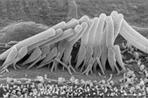
Corentin Affortit
College of Medicine
Otolaryngology – Molecular Otolaryngology & Renal Research Laboratories
Category: Faculty/Staff/Researcher
Our lab research focuses on discovering new genes associated with genetic deafness and to decipher the associated mechanisms.
This picture shows a high magnification image of a mouse cochlear inner hair cell using scanning electron microscopy. The inner hair cells are the primary sensory receptors in the cochlea, responsible for converting mechanical sound vibrations into neural signals. In this high magnification picture, only the apical surface of the inner hair cell is visible, showing the unique architecture of their hair bundles. This precise arrangement of the stereocilia is crucial for their function and alteration of these hair bundles such as missing, fused, or elongated stereocilia is a common cause of sensorineural hearing loss. Delicate tip-link connections essential for mechanoelectrical transduction may be seen between the tips of shorter stereocilia and the sides of taller ones.
Better understanding of the morphological alteration induced by gene mutations is very valuable in appreciating the mechanism associated with genetic hearing loss.
Acknowledgements: Dr. Richard Smith
Bloodshot Clot Plot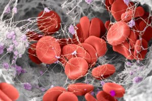
Milad Arzani
College of Engineering
Biomedical Engineering – BioMOST
Category: Graduate
Colored scanning electron microscope (SEM) image of a human blood clot. Clot is a double-edged sword. It is vital to life because it is what stops bleeding, while on the other hand, it can also disrupt life if it occurs at the wrong place and time. I study stroke, which occurs when a blood clot blocks an artery in the brain. If it isn’t removed quickly, patients can lose bodily functions for good. But some clots are easy to remove, and others aren’t. Why? I want to understand what makes up a clot. How mechanically coherent is it? Are there different phenotypes in structure and coherence? Can we infer these phenotypes through diagnostic imaging? Questions at the interface of biology, mechanics, and imaging that engineers are well-placed to ask and answer. Answers that will lead us to better treatment selection and improved outcomes for stroke patients worldwide. This clot was surgically retrieved from a stroke patient’s brain. It showcases the microscopic structure of blood clots, revealing a complex tapestry of red blood cells enmeshed within a dense fibrin network, interspersed with small platelet aggregates.
Acknowledgements: I want to express my gratitude to Professor Raghavan and Professor Ortega for their guidance and support throughout the project, as well as to Dr. Cruz for his contributions, and Thomas Moninger at CMRF for his help with the biological sample preparation for SEM.
Phase-Locked Loop Frequency Response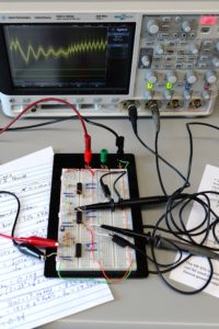
Kara Beauchamp
College of Engineering
Electrical and Computer Engineering – Departmental Undergraduate Education
Category: Faculty/Staff/Researcher
The image shows an analog electronic circuit called a phase-locked loop built out on a breadboard along with an oscilloscope displaying two time-varying signals from the phase-locked loop. A phase-locked loop is a type of electronic control system that adjusts the output signal of an oscillator to match the frequency and phase of an input signal through the use of an intermediary signal called the tune voltage. Phase-locked loops are essential in many technologies, including radios, cell phones, GPS receivers, and computers, where they help synchronize signals and recover timing information.
The signals shown on the oscilloscope are the input to the phase-locked loop (shown in green) and the tune voltage of the phase-locked loop (shown in yellow). The input is a square wave which abruptly changes in frequency from higher to lower (on the left side of the oscilloscope trace) and back to higher (on the right side of the oscilloscope trace). The yellow signal shows the response of the tune voltage to the change in the input frequency.
In an ideal phase-locked loop, the tune voltage would instantly respond to the change in frequency by dropping from high to low voltage when the frequency dropped from high to low and then rising from low to high when the frequency rose. This phase-locked loop is not designed for a fast response to the change in input frequency and it creates a more interesting-looking signal. When the frequency changes from high to low, the tune voltage experiences “ringing” where the tune voltage both overshoots and undershoots the desired low voltage level. In the pictured case, the tune voltage has a form which is similar to the oscillation amplitude of an underdamped oscillating mass on a spring, demonstrating analogous behavior between a mechanical system and an electronic system
Acknowledgements: Anton Kruger suggested the design, Phil Thompson created the detailed design, and Hailey Boileau took the picture.
Cells in Orbit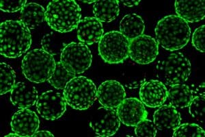
Emily Formella
College of Engineering
Biomedical Engineering – Worthington Lab
Category: Undergraduate
This confocal microscopy image shows retinal pigmented epithelium cells adhered to spherical microcarrier beads, visualized using fluorescent viability staining (green = Calcein AM for live cells; red = Ethidium Homodimer for dead cells). Culturing these cells on microcarriers (average diameter of 150 μm) allows them to be maintained in a rotating bioreactor suspension culture, which serves as a ground-based model to simulate certain mechanical aspects of microgravity.
This research investigates how simulated microgravity influences chorioretinal cell behavior, focusing on mechanotransduction pathways that decide cellular structure and function. By studying these responses, we stand to better understand the visual changes that occur outside of Earth’s atmosphere and the health risks posed to long-duration astronauts.
Acknowledgements: I would like to thank Dr. Worthington for providing this exciting research opportunity and all members of her lab for their continued support.
Tracking Life Through Painted Shells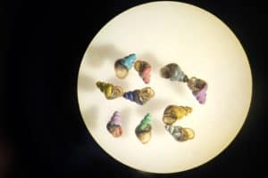
Alejandra García-López
College of Liberal Arts and Sciences
Biology – Neiman Lab
Category: Faculty/Staff/Researcher
This image, taken with a binocular microscope, shows a group of aquatic snails of the species Potamopyrgus antipodarum, commonly known as the New Zealand mud snail. These tiny mollusks, measuring only 2 to 5 millimeters in length, serve as an ideal model for investigating fundamental ecological questions such as the maintenance of sexual reproduction and population dynamics. They are also widely used in ecotoxicological studies and are particularly relevant for being among the most widespread invasive aquatic species worldwide.
Working with organisms so small presents significant challenges. To facilitate their monitoring, we employ a simple yet ingenious method: painting each shell with non-toxic acrylic paint. Each color becomes a unique “barcode,” allowing us to track specific individuals over time. This approach is especially useful for monitoring individual growth under different conditions, distinguishing males and females in mate-choice experiments, and identifying the lineage of each specimen.
The precision required to work with these millimetric animals demands both patience and precision—skills that are refined day by day in the laboratory while measuring, painting, sexing, photographing, and dissecting them. This image embodies the dedication behind uncovering key ecological questions and deepening our understanding of biological invasions, transforming a tiny mollusk into a remarkable source of scientific insight. This image represents the dedication to unravel key ecological questions and to better understand biological invasions, transforming a tiny mollusk into a remarkable source of scientific knowledge.
Acknowledgements: Maurine Neiman (all members of Neiman Lab), International Programs, Ana R.R.
Going With The Flow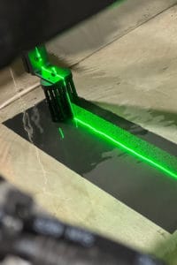
Abhishek Ghimire
College of Engineering
Civil and Environmental Engineering – IIHR
Category: Graduate
Leonardo da Vinci, in his famous sketch from 1510, gave one of the earliest explanations of fluid flow and random rotational motions in flow called “turbulent eddies”, simply through observation of flowing water using the naked eye. These turbulent eddies affect the flow of energy and things like sediments in the flow. Fast forward to present day, we now have different techniques to visualize precisely how fluid moves in nature and how energy flows along with it. One of such techniques is called Particle Image Velocimetry (PIV), which is used to observe and study fluid flow by illuminating particles in the fluid with a laser while capturing images at a very high rate. This picture shows the use of PIV to study the flow field behavior when water flows through a patch of vegetation in a laboratory. This can be extrapolated to understand the flow behavior around vegetation in nature and the potential use of vegetation in the management of water channels and as a nature-based solution against erosion.
Acknowledgements: Dr. Priscilla Williams
TRACERS, Dr. Craig Kletzing’s Legacy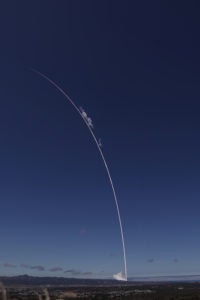
Christian Hansen
College of Liberal Arts and Sciences
Physics and Astronomy
Category: Faculty/Staff/Researcher
On July 23rd, 2025, a SpaceX Falcon 9 launched the TRACERS mission into orbit, capping off an eight-year effort from mission concept, design, build, test, to finally on orbit. TRACERS, the University’s largest ever single award, is a NASA funded mission led by the Department of Physics and Astronomy to deliver a pair of identical satellites each hosting a suite of science instruments to study magnetic reconnection and its effects on Earth’s atmosphere. TRACERS was initially led by Dr. Craig Kletzing and was the culmination of Dr. Kletzing’s career but sadly he passed away before launch. Dr. David Miles took over for Dr. Kletzing as his health was declining and led the team to the finish line. To commemorate Dr. Kletzing’s legacy, one of his personal guitar picks was embedded into the science computer on each satellite. The TRACERS satellites and the researchers involved work to carry out his legacy through scientific discovery. The launch streak is a composite image of thousands of video frames stacked together to produce one single streak that shows the rocket’s trajectory as it races towards low earth orbit. The video was taken 12 miles away from the launch site and the rocket at the end of the streak is at an altitude of 36 miles.
How to Train your Biomarker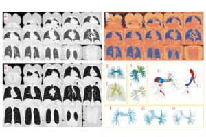
Soheil Hosseini
College of Engineering
Biomedical Engineering – APPIL
Category: Graduate
This vibrant mosaic captures the journey of a new AI-based biomarker—from the grayscale world of clinical CT scans to the colorful geometry of data-driven discovery. Each quadrant tells a chapter in the biomarker’s creation: the quiet rhythm of coronal lung slices at rest and in full inspiration on the left; the vivid interplay of image registration aligning the lungs across the two volumes at top right; and finally, the emergence of translucent 3D models—airways glowing in gold, arteries shimmering in blue, lungs breathing in greens.
Beneath this beauty lies precision. Artificial intelligence, trained on expert knowledge, learns to trace the hidden harmony between airways and arteries, identifying 24 standard locations linking airway and arterial anatomy that define our new vascular biomarker, ArtLR—the Arterial-to-Lung Ratio. Here, art and science meet: transparent branches reveal the intricate graphs and nodes that define the shape of human breathing, while color illuminates the matched branches, showing structure and scale in vivid clarity.
The image is both map and metaphor—a portrait of the lung as living architecture, where every pixel reflects precision, data, and discovery. It invites viewers to see how mathematics, medicine, and engineering converge to deepen our understanding of disease—to disentangle the intertwined contributions of airways and arteries and bring us closer to uncovering the origin(s) of COPD susceptibility and pathogenesis.
Acknowledgements: Prof Eric A. Hoffman
Tracking Bur Oak Blight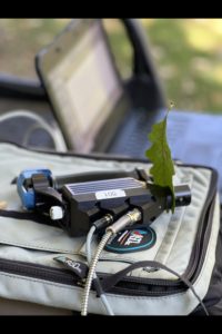
Mainul Islam
College of Liberal Arts and Sciences
School of Earth, Environment, and Sustainability – LOQATE
Category: Graduate
Bur Oak Blight is a fungal disease in Bur Oak leaves. Leaves are the primary way to check the existence of blight. In this picture, I am using a Spectrometer setup to collect leaf spectral signatures. Blighted leaves have specific signals compared to healthy leaves. I am tracking the signals in the nature of Iowa!
Acknowledgements: Dr. Susan Meerdink, Dr. Mary Skopec, Nora Gibson, Alex Arens
In the Weeds with Flood Forecasting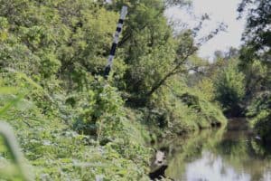
Anthony Lamoreux
College of Engineering
Civil and Environmental Engineering – IIHR
Category: Graduate
Flood forecasts depend on static river models that are calibrated for the upstream rivers of a town or city, but studies have found a substantial increase in predicted maximum flood depth when using a dynamic river model over a static one. The difference between dynamic models versus a static model is that they consider the plant height on the riverbanks which is shown in this image. Grasses starting out at a few inches tall in the spring bloom into a towering 6-foot-tall plant in the early and late summer. This drastic change in height measured by my striped yellow and black poles leads to a 20% increase in river resistance, thus slowing down the river more than the static model anticipated. An effect of slowing down a river is increasing the depth of the river; therefore, flood forecasts using the dynamic river model are 7% more accurate in maximum flood peak timing and depth. This result helps improve public and private property flood preparation where resources and time are already limited.
Acknowledgements: Dr. Marian Muste
2025 Voice Foundation Symposium Program Cover Image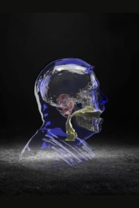
David Meyer
College of Liberal Arts and Sciences
School of Music & UI Communication Sciences and Disorders
Category: Faculty/Staff/Researcher
This image is a compilation of MRI scans of my ear, vocal tract (the airspace in the throat – from my vocal folds to lips), skull, and the external surface of my head. I am singing an /a/ vowel as in the word, “father” in an operatic voice. Singers are the only musicians whose instrument is invisible. At the University of Iowa, we are using medical imaging in an interdisciplinary collaboration with Biomedical Engineering and Radiology, to better model the vocal instrument. MRI is the safest modality (as compared to CT), but it does not capture bone. The skull in this image is the result of a new imaging technique in MRI, zero echo time MRI (ZTE-MRI), further optimized here at the University of Iowa (with collaborations with GE HealthCare), and Mayo clinic in Minnesota. This allows us to safely ‘see’ bone in MR, free from exposure to ionizing radiation (common in x-rays and CT). This image was selected by the Voice Foundation to be the cover of their international symposium in Philadelphia last summer (2025).
Acknowledgements: Three of Dr. David Meyer’s interdisciplinary collaborators were instrumental in the generation of this image: Dr. Sajan Lingala (Biomedical Engineering faculty) his lab (graduate student: Swati Ramtilak), and Ian DeNolfo (Executive Director, Voice Foundation).
Last Look at the TRACERS Satellites on Earth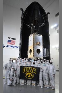
David Miles
College of Liberal Arts and Science
Physics and Astronomy – Iowa Spaceflight Laboratory
Category: Faculty/Staff/Researcher
The University of Iowa is investigating the mysterious and powerful interactions between the magnetic fields of the Sun and Earth. The TRACERS (Tandem Reconnection and Cusp Electrodynamics Reconnaissance Satellites), consisting of two identical satellites orbiting Earth in tandem (one following the other). They will help answer long-standing questions key to understanding space weather, particularly how the Sun transfers energy, mass, and momentum to near-Earth space. The two satellites are shown in their final state on Earth. After this photo, two halves of a protective fairing were closed over the satellites, they were mounted on a SpaceX Falcon 9 rocket, and on July 23, 2025 they were successfully launched into space.
Acknowledgements: SpaceX and NASA Kennedy Space Center
It’s all about the MAC!
Rachel Moore
Carver College of Medicine
Neuroscience and Pharmacology – Mulfual Lab
Category: Graduate
Human induced pluripotent stem cells (hiPSC’s) are differentiated into choroidal endothelial cells (CEC’s). Once they create a monolayer, the cells are injured by complement normal human serum to form the Membrane attack complex (MAC) on the cells. This picture depicts the CEC’s by a tight junction protein CDH5 in green, and MAC formation on injured cells in red.
Acknowledgements: Kelly Mulfaul
Where Light Meets Precision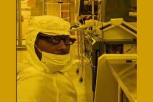
Arinze Okoye
College of Engineering
Mechanical Engineering/Physics – Prineas Research Group
Category: Graduate
Inside the golden glow of a photolithography lab, precision and patience converge. This image captures a defining moment in semiconductor fabrication—where light patterns are transferred onto microscopic wafers to shape the foundation of next-generation devices. Wearing a full cleanroom suit, I stand amid the quiet hum of vacuum pumps and alignment systems, where even a speck of dust can alter a nanometer-scale design.
My research focuses on improving infrared photonic devices and detector architectures through advanced photolithography and etching techniques. Each wafer processed in this room represents countless experiments to optimize light absorption, enhance quantum efficiency, and refine surface morphology. The yellow illumination symbolizes the filtered environment that protects photoresist materials from premature exposure—an unseen safeguard in every successful pattern transfer.
This image reflects both the artistry and discipline of microfabrication: a space where human intent interacts with light, chemistry, and matter to define the invisible structures that power modern technology. It is a reminder that innovation often begins in the quiet, golden stillness of the cleanroom.
Acknowledgements: Prof. John Prineas
Hawkeyes in the Skies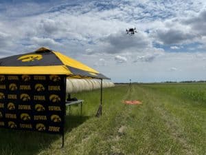
Jalissa Pirro
College of Liberal Arts and Sciences
School of Earth, Environment, and Sustainability – LOQATE
Category: Graduate
In an age where advancing technology brings forth new and exciting opportunities in research, Unpiloted Aerial Systems (UAS) enable scientists to take to the skies and study the environment in new ways. In collaboration with Iowa State University’s Agronomy Department, I use UAS-mounted Light Detection and Ranging (LiDAR) to study Miscanthus × giganteus (MxG), a perennial tall grass that grows up to twelve feet and is converted into biomass fuel. Taken during a late-May flight campaign, my photo shows a DJI Matrice 300 RTK capturing LiDAR imagery that will be used to map MxG height and distribution across the adjacent field. Since MxG is not replanted every year, understanding plant survival and identifying areas of low productivity in the field are essential for guiding management strategies. Through my research, I aim to advance the use of UAS for understanding long-term crop productivity and to provide data that supports management strategies for maintaining high and sustainable yields.
Acknowledgements: Susan Meerdink and Samuel Taylor
Quantum Cascade Laser Light Test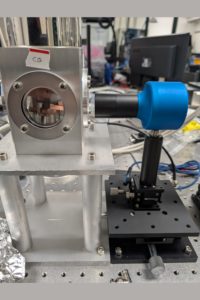
Adam Ramker
College of Engineering
Electrical and Computer Engineering – Toor Lab
Category: Graduate
Left: a cryostat—an insulated chamber that we evacuate (remove air) and cool to near absolute zero so heat and moisture don’t disrupt sensitive measurements. Right: a pyroelectric light-power detector. It measures optical power by absorbing light, turning that energy into heat, causing a special crystal to expand and contract (thermal vibration), which generates a voltage proportional to the light’s power.
Inside the cryostat sits the star of the shot: a semiconductor quantum cascade laser (QCL). Unlike ordinary lasers that rely on electron–hole recombination, a QCL uses carefully stacked quantum wells to “cascade” electrons down energy steps, emitting photons at each step. This design lets us reach wavelengths that are otherwise hard to access.
This image captures the laser’s first run at 10 Kelvin (−263.15 °C). At that temperature, random thermal noise drops and the device’s internal states sharpen, so tiny details in its behavior become visible. The pyroelectric detector on the right closes the loop—converting the laser’s invisible output into a clean electrical signal we can analyze. Cold, quiet, and precise: this is what it looks like when quantum engineering meets a simple question—“How much light is really there?”
Acknowledgements: Dr. Fatima Toor
The Smart Field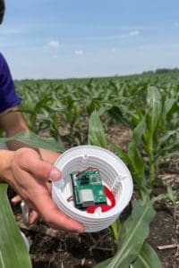
Rafael Rangel de la Tejera
College of Engineering
Chemical and Biochemical Engineering – Atmospheric and Environmental Research Lab
Category: Graduate
This image embodies the essence of my research on developing and deploying “Internet of Things” based smart sensors that monitor microclimate conditions in agricultural fields. The sensor shown here, held among young corn plants, represents the intersection of technology, environment and innovation. After this photo was taken, the sensor was installed in the middle of the field to record temperature, humidity, air pressure, and soil data, transmitting it in real time for farmers to access and visualize field conditions. This deployment took place at Schott Family Farms in Riverside, Iowa, as part of a broader initiative to establish a sensor network across multiple Midwestern sites for continuous environmental monitoring. The image is meant to represent how technology can be applied to strengthen agricultural resilience and improve decision making, bridging the gap between science and sustainable farming practices.
Acknowledgements: Dr. Jun Wang and Dr. Lorena Castro
In Vivo Bone Micromechanics of the Human Distal Tibia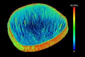
Punam Saha
College of Engineering
Electrical and Computer Engineering
Category: Faculty/Staff/Researcher
Stress distribution along bone microstructures in the human distal tibia, assessed using high-resolution peripheral quantitative CT (HR-pQCT) in vivo imaging and a finite element analysis (FEA) algorithm incorporating nonlinear continuum mechanical modeling, pioneered by our research team. A unique feature of this method is its ability to quantify bone micromechanics without requiring segmentation of bone microstructures. This relaxation of binary segmentation extends the method’s applicability, enabling quantification of micromechanical properties while capturing bone microstructural distribution at the scanner’s intrinsic resolution. The image illustrates stress flow lines along the trabecular bone network without stress leakage into the marrow space, demonstrating the method’s effectiveness in capturing bone micromechanical behavior. Regions along the cortical bone rim show high stress concentrations, supporting anatomical knowledge that cortical bone plays a primary role in bearing body weight and mechanical loads during normal physical activity.
Acknowledgements: Xiaoliu Zhang
Mutual Support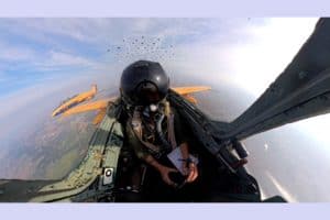
Tom Schnell
College of Engineering
Industrial and Systems Engineering – ITI
Category: Faculty/Staff/Researcher
Two research jets of the Operator Performance Laboratory (OPL) are heading out on a USAF-funded research mission in fingertip formation. Military aircraft operate in tactical formations to provide mutual support and to conduct the mission more effectively. Flying in formation requires a fine touch and the pilots must operate predictably and with precision. Trust in the actions of the other pilot is essential and must be earned. Once the formation arrives in the Area of Operation, it will spread out and adapt to tactical requirements. At that point, the aircraft may operate many miles apart but, through a net-centric connection, still move in close coordination to provide mutual support, similar to a wolf pack. This mission involves testing autonomous autopilot algorithms that give military aircraft the ability to operate tactically without pilot input. This frees up cognitive resources of the pilot. We measure cognitive workload using an ECG based method. We quantify pilot attention using an eye tracking system that is integrated into the helmet seen in the picture. Under the helmet visor is a small optical combiner that provides the pilot with flight and mission-essential information. Onboard are many computers that aggregate incoming information in real-time and provide the autonomy system with control commands. Shortly after this picture was taken, the two jets were fighting against incoming cruise missiles that are generated in a simulation on the ground, and which are transmitted as tracks to the mission computers on the jets. This work, which is a national defense priority, supports graduate students who are active-duty USAF Test Pilot School (TPS) graduates and who are in the process of earning their Ph.D degrees at OPL under the mentorship of Professor Tom “Mach” Schnell.
Acknowledgements: USAF Test Pilot School
Bridge Sensor Assemble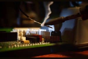
Mia Smith
College of Engineering
Civil and Environmental Engineering – IIHR
Category: Undergraduate
The Iowa Flood Center (IFC) installs flood sensors on bridges across the state to give communities early warnings and time to prepare for rising waters, especially along rural roads. Each solar-powered sensor is a custom built assembly of parts, sensors, and modems. Their modular design allows for quick repairs and upgrades as technology changes. Some components require precise soldering and careful assembly to ensure durability and accurate data transmission in Iowa’s varying weather conditions. This image shows the hands-on work behind each sensor and highlights the IFC’s effort to build reliable tools that help keep Iowa’s communities safe from floods.
Acknowledgements: James Niemeier
The Smiling Sun: When the Star Showed Its Joy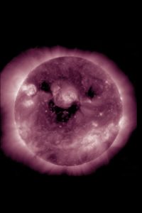
Shirsh Soni
College of Liberal Arts and Sciences
Physics and Astronomy – Miles Lab
Category: Faculty/Staff/Researcher
On October 26, 2022, NASA’s Solar Dynamics Observatory (SDO) captured a rare and delightful sight, a “Smiley Sun.” In this ultraviolet image, taken by the Atmospheric Imaging Assembly (AIA) at 211 Ångström, the dark regions on the solar disk appear to form two eyes and a curved mouth, giving our star an unexpectedly human expression. These darker patches are known as coronal holes — areas where the Sun’s magnetic field opens into space, allowing high-speed solar wind to escape into the heliosphere.
Beyond its cheerful resemblance, this image is a powerful reminder of how science and nature often merge in beauty. The patterns we see are the result of complex magnetic activity and plasma dynamics on the Sun’s surface, yet they evoke emotions that transcend data and numbers. This moment of cosmic coincidence, the Sun smiling down upon us, resonated worldwide, reminding humanity that even the most dynamic and powerful forces of the universe can show a gentle face.
For me, this photograph symbolizes the harmony between science and wonder. It illustrates how observation and imagination coexist, how a satellite’s precise instruments can capture not just phenomena, but personality. The small blue dot in the upper corner shows the scale of Earth, placing our tiny world in perspective against the vast, radiant smile of the Sun.
In a time when we often look to space for answers, this image whispers a simple truth: the universe, too, has its moments of joy.
Acknowledgements: NASA’s Solar Dynamic Observatory Team
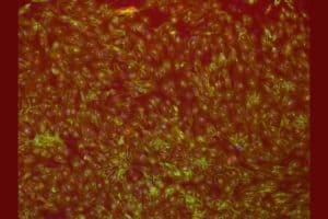
Drosophila Melanogaster Ovarian Follicle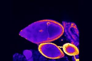
Sean Strand
College of Liberal Arts and Sciences
Biology – Tootle Lab
Category: Undergraduate
This image is of a Drosophila Melanogaster (fruit fly) ovary that has been stained with various primary and secondary antibodies and DNA stains to observe structures under immunofluorescence microscopy. This specific image acts as the control for the experiment being tested. The inner cluster of cells are a model for cluster cell migration, a cell movement pattern not fully understood. Understanding how clusters of cells move and how they communicate with one another can provide insight into cancer cell metastasis, and it’s overall prevention. The cells on the outer sides of the follicle act as the determinants of timely cell migration, or reaching the egg at the same time the outer follicle cells do. By looking at this follicle which is in stage 9 of it’s maturation we can determine through comparisons of outer follicle cells and inner cluster cells that migration is on time for our control group.
Acknowledgements: My Graduate student and mentor, Ashley Goll, and our Principal Investigator, Dr. Tina Tootle
Honey, I Shrunk the Tumors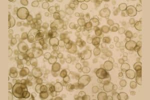
Hiruni Sumanasiri
College of Liberal Arts and Sciences
Microbiology & Research in OBGYN – Kristina Thiel Lab
Category: Undergraduate
At first glance, this image looks like floating soap bubbles, but these are actually organoids grown from healthy endometrial tissue. Organoids are tiny 3D clusters of cells that recapitulate the structure and function of human tissue on a miniature scale. In the Thiel lab, we grow both healthy and cancerous organoids to study gynecologic cancers such as endometrial and ovarian cancer.
Although the organoids that are in this image are non-cancerous, they still play an essential role in our research. Healthy models act as the baseline, helping us understand the difference how cancerous cells differ in growth, shape, as well as response to treatment. In “shrinking” these complex tissues into bubble like structure, we can study cancer in a controlled environment.
Acknowledgements: I would like to thank my graduate student Kaitriana Colling!
Iowa as Above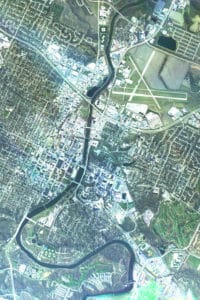
Star Tammes
College of Engineering
Chemical and Biochemical Engineering – AER lab
Category: Graduate
The NIGHTHAWK prototype airborne imager is a collaborative development project involving multiple departments across the university, supported through funding by the P3 program. Designed as an experimental imaging system, NIGHTHAWK is capable of capturing separate red, green, and blue (RGB) spectral bands, as well as a NIR channel not shown, while mounted on a light propeller-driven aircraft owned by the OPL. During operation, the system records images in rapid succession as the aircraft passes over the target area at altitudes of a few thousand feet. These individual RGB images are then computationally aligned and merged, or stacked, to create true-color composite mosaics. This process produces a seamless, high-resolution view of both the town and the university campus, revealing fine spatial detail that would not be possible with single-band photography.
The flight documented here took place on November 26th, 2024, and served as one of the early test campaigns for the NIGHTHAWK platform. The aircraft completed several circuits around town, flying at three distinct altitudes in order to evaluate how well the imaging and alignment algorithms performed under varying conditions. This test was a critical step in validating the system’s ability to compensate for differences in perspective, scale, and motion. The results demonstrated the feasibility of the approach and provided a proof of concept for using low-cost aerial imaging systems in future research and applied projects.
Acknowledgements: Dr. Marc Linderman, Dr. Tom Schnell, Dr. Jun Wang
The Loneliest Generation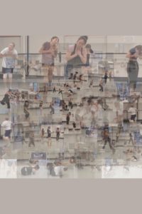
Christina Yu
College of Liberal Arts and Sciences
Dance
Category: Undergraduate
Take a look at the directed gazes, angles, and emotions present in the dancers, and notice the lack of interaction between each individual and the collective.
As people, we long for real connection—connection that goes beyond digital barriers, highlights, and spectacles. And yet, our lives continue to revolve around this “social” media world. Shown in this collage of images is a digital diary of eight dancers experimenting with this paradox through dance: how can one generation be so connected and yet feel so alone? This paradox embodies the vision of Lágrimas do Paraíso, a dance created by Professor Eloy Barragán to magnify and display these feelings of collective isolation—moving together, yet separate. As one of the eight dancers, my role is to bring these conflicting ideas to life through movement, whether onstage or off. This collage I created is an amalgamation of curated screenshots sourced from a single rehearsal video. Its square format and pixelated quality are meant to leave you with a sense of irony—that even now, this art was created through a screen.
Acknowledgements: Thank you to Eloy Barragán for including me in your special process. Another thank you to Victoria Adams, Iliana Banu, Andi Bartlein, Payton Conrad, Brenna Labus, Natalie Prill, and MJ Van Ostrand for being such great friends and dancers for this project. We did this together.