
Voting is now closed!
The Silky Horizon
Milad Arzani
College of Engineering
Biomedical Engineering – Mu Lab
Category: Graduate Student
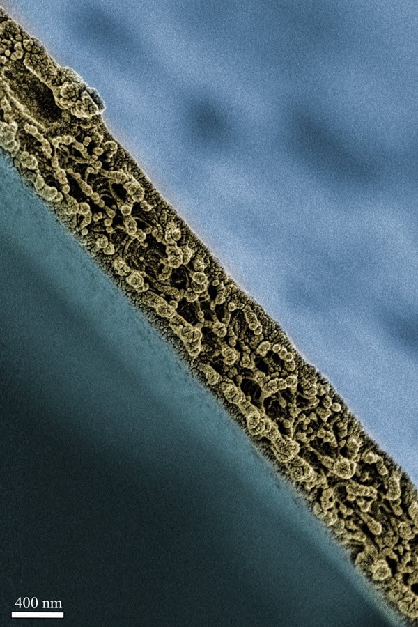
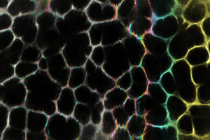
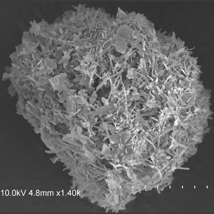
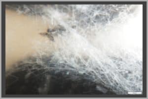
Captured at the Central Microscopy Research Facility at the University of Iowa, this cross-sectional scanning electron microscope (SEM) image reveals the intricate nanogranular structures within a thin silk fibroin film of a thickness of around 300 nm, roughly one three-hundredth of the diameter of a human hair (100 µm). The intricating nanostructures imply the potential assembling pathway of silk fibroin molecules, which underlies the spin coating-assisted fabrication. More interestingly, the film is made of 100% proteins, the same building blocks of our bodies. Due to the unique combination of proteinaceous composition and thinness at several hundred nanometers, this film will be advantageous in constructing essential tissue barriers, controlling membrane-mediated mass transport, and interfacing wearable and implantable biomedical devices, thus exhibiting new horizons in various biomedical applications. The SEM image is colored in UIowa gold to highlight the thinness of the silk fibroin film.
Acknowledgments: I want to thank my advisor, Professor Xuan Mu, for his support and patience and for allowing me to learn SEM imaging.
 Accessible Pharmacology
Katelin Dannen
Carver College of Medicine
Neuroscience and Pharmacology
Category: Faculty/Staff/Researchers
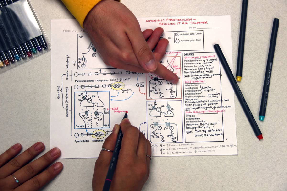
Pharmacology (how drugs work) is a challenging subject requiring students to mentally overlay complex mechanisms of drug action onto intricate body physiology to make sense of how drugs both correct and cause disease. Despite this challenge, limited research has been done to define the most effective pharmacology pedagogy. Therefore, Dr. Dannen has developed critical thinking exercises and guided diagramming activities to break up the often lecture-heavy pharmacology curriculum in her undergraduate courses. Furthermore, she is assessing whether these collaborative, active learning strategies make Pharmacology material more understandable and accessible to students.
Dr. Dannen also recognizes that, in addition to its critical role in healthcare curriculums, Pharmacology may also benefit students whose professions lie outside of healthcare. Drugs, both therapeutic and non-therapeutic, are common on college campuses. Access to Pharmacology education in college may serve to create a culture of more educated consumers and to reduce drug misuse. Therefore, Dr. Dannen is also measuring the extent to which elementary and advanced undergraduate Pharmacology coursework influences student drug consumer knowledge and use.
This photograph captures students collaborating on a guided diagraming activity to bring together 4 weeks of Pharmacology class material into a one-page study guide.
Dannen KE, Critical Thinking Pharmacology Worksheets. Iowa Research Online (ISSN 2476-1680). 2023
Acknowledgments: I would like to acknowledge Madelin Schwager and Joshua Santiago, two students in Pharmacology I (PCOL:3101), who agreed to pose for the picture. I would also like to acknowledge Linda Buckner for taking the photograph.
 Algae Thrives in Wastewater
Halle Eisfelder
College of Engineering
Environmental Engineering – IIHR – Hydroscience & Engineering, Just Research Group
Category: Undergraduate Student
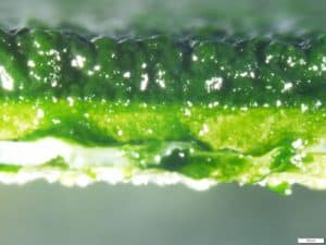
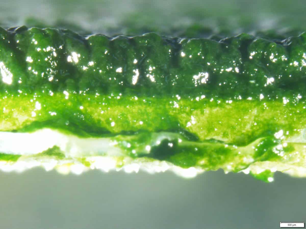
The algae in this photo were grown in the lab using rotating algal biofilm (RAB) units by attaching cotton material to a microscope slide. The slide was submerged in a mixture containing high strength wastewater, a pH buffer, and water. The algae biofilm utilizes the nutrients in the wastewater to grow thick and fluffy. This alga could potentially be land applied as fertilizer since it assimilates the nitrogen and phosphorus in the wastewater. The thickness of the algae is highlighted in this photo and is directly related to the ammonia loading. My research hopes to demonstrate that algae biofilms behave like other biofilms used in wastewater treatment. The Monod Kinetics demonstrated in this experiment are key to understanding the relationship between algae thickness and nutrients. The image was captured using the Olympic Stereoscope microscope. This research overall allows for better understanding and future application of algae in treating wastewater.
Acknowledgments: Maclaine Putney and Craig Just
 Iowa SUDEP Research Program
Sofia Espinoza
College of Engineering
Neurology – Neuroscience Laboratory Department
Category: Undergraduate Student
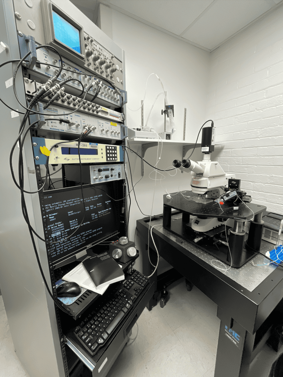
The photograph captures my research station at work in a neurology lab, dedicated to studying sudden unexpected death in epilepsy (SUDEP). In the image, the computer screen displays EEG, EKG, and neuronal membrane potential recordings from a mouse model. Various electrodes and sensors are attached, measuring critical parameters like the excitation of neurons (arousal), oxygen and carbon saturation, and pH. The written computer code is to process and analyze the data, with a setup of signal conditioning hardware visible in the background. My role involves coding programs and methods for precise data collection and conducting experiments aimed at understanding the mechanisms behind SUDEP, possibly drawing parallels to sudden infant death syndrome (SIDS). This hands-on work is a vital part of a broader research effort, contributing to potential life-saving insights in the field of epilepsy and neurology.
Acknowledgments: Dr. Eduardo Bravo, Dr. George Richerson, Dr. Frida Teran-Garza, Dr. Karina Bravo, and Mr. Xiuqiong Zhou
 Research Companions
Allison Flores
College of Engineering
Electrical and Computer Engineer
Category: Graduate Student
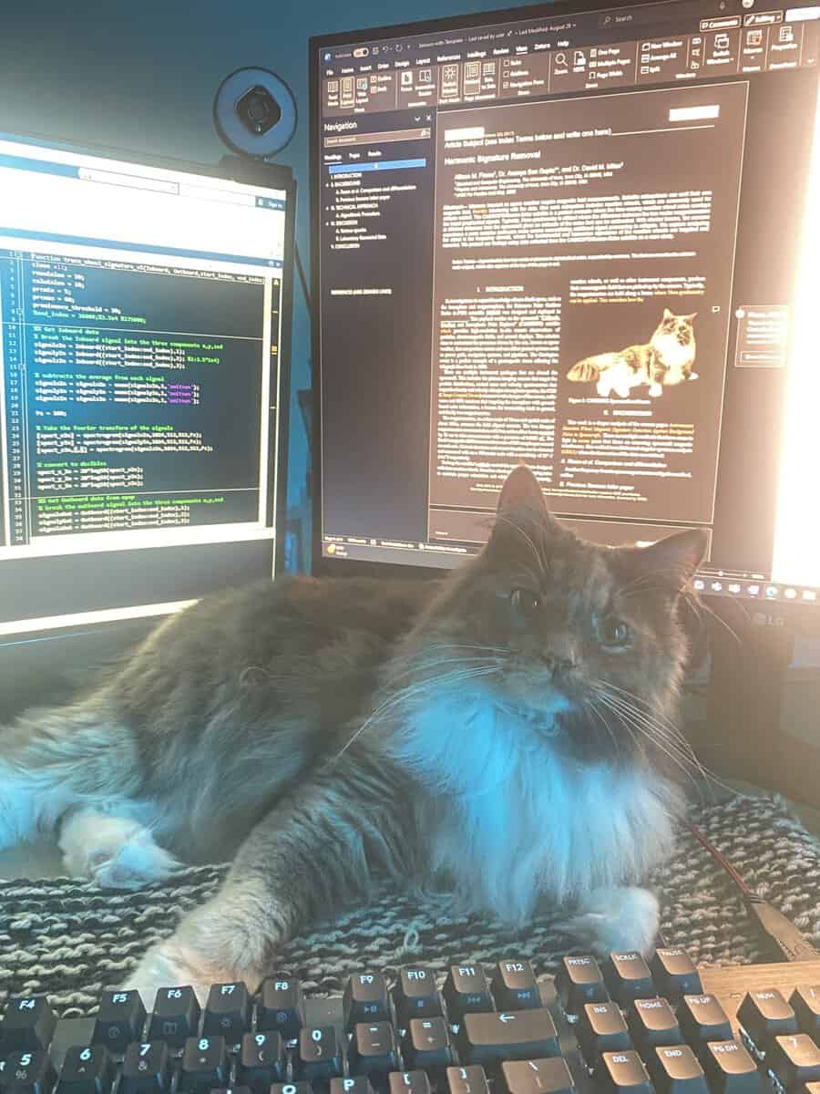
In this photo, I’m joined by my loyal assistant, Birdie, as we tackle late-night research sessions. Here I’m working on code for my journal paper under the guidance of my advisors, Dr. David Miles and Dr. Ananya Sen Gupta. My work focuses on developing signal processing algorithms to isolate magnetic interference in spacecraft data. Late nights like this, with Birdie by my side, are all part of the journey through the PhD grind– tackling complex problems and pushing the boundaries of knowledge one line of code at a time.
Archives Rat
Max Halbach
College of Liberal Arts and Sciences
History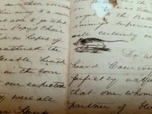
Category: Undergraduate Student
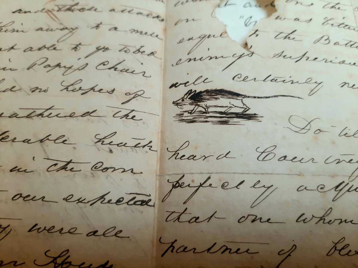
The photo comes from a letter found in the University of Virginia’s Albert & Shirley Small Special Collections Library. The letter was written (and drawn) by the brother of Thomas H. Bakken on October 31, 1841 (Halloween). Thomas’ brother lived in Louisiana, and he wrote back to his Virginian family about life in the Old Southwest. In this letter, he complains about how rats frequently eat his crops, and the lengths he goes to try to stop them. Interrupting his story, he draws a small whiskered rat with a dark back scurrying over his cursive writing. A hole near the top of the letter makes the observer wonder if it was simply age that created it or the leftovers of another rat’s meal. Marginalia, or margin art, is rarely featured in popular history books, yet it is a form of art that can reveal concerns about westward expansion in the 1840s; did he include it to entertain or frighten? As a reference or a warning?
Acknowledgments: The University of Iowa Department of History for their generous funding allowing me to travel to, and research at the University of Virginia
 Lost in Vibration
Jacob Herrmann
College of Engineering
Biomedical Engineering
Category: Faculty/Staff/Researchers
Our research focuses on microstructural dynamics of lung tissue, at the alveolar scale where gas exchange occurs. In this image, a sample of lung tissue is mechanically vibrating while being scanned by a confocal microscope. The laser scans columns of pixel data vertically, from top to bottom across the image. Lung tissue is autofluorescent and emits light when excited by the laser, while the gas contained between the alveolar walls remains dark. Each grayscale pixel has been pseudo-colored according to the corresponding phase of the periodic vibration cycle. The vibrating tissue appears distorted depending on the speed of the scanning laser relative to the vibration frequency. From left to right across the image, the relative speed of the scanning laser is accelerated. On the left, scanning is so slow that multiple vibration cycles occur within each column, and only an average sense of structure is apparent, with the colors blending to appear gray from far away. In the middle, each column represents a distinct phase, and the image appears wavy. On the right, scanning is so fast that the vibrating sample appears stationary (i.e., a single phase or color).
Acknowledgments: Daniel Meggo, Edward Sander
 BioHawkeye: Mastering the Art of Protein-Protein Interface Predictions
Klaertje Hesselink
College of Engineering
Chemical and Biochemical Engineering – Gomes Lab
Category: Undergraduate Student
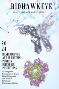
The cover art for my theoretical journal, BioHawkeye, represents the intersection of biological concepts and machine learning, focusing on research conducted in the Gomes Lab at the University of Iowa. This picture was created using Canva and Visual Molecular Dynamics (VMD), a molecular visualization program for displaying large biomolecular systems using 3-D graphics and built-in scripting, where this design reflects a modern approach to scientific visualization. In this journal cover, two protein structures are depicted through ribbons (which represent the primary sequence of the protein), spheres, and black lines. The black lines represent connections between alpha carbons (spheres), illustrating potential interactions within a protein. Surrounding the proteins are vibrant dots and lines, symbolizing the principles behind Graph Neural Networks, highlighting how these computational tools help analyze and predict protein interface changes. This design emphasizes the role of technology in uncovering patterns in biological data, inviting readers to explore innovative methods for understanding proteins. The research featured in BioHawkeye primarily focuses on developing machine learning models to predict protein-protein interface prediction which is an ongoing project in the Gomes Laboratory.
Acknowledgements: I want to thank the UI-MARC fellowship program for funding this research and Dr. Joe Gomes for mentorship and support.
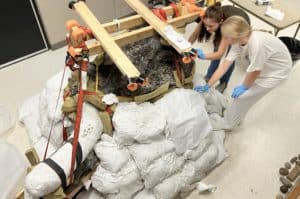 Maya and the Mastodon
Tiffany Adrain
College of Liberal Arts
Earth and Environmental Sciences – Paleontology Repository
Category: Faculty/Staff/Researchers
UI undergraduate and OUR Research Fellow, Maya Monk (biology/environmental science major), conserves a 13,600-year-old mastodon skull with assistance from Seraphina Carey at the Office of the State Archaeologist (OSA). Maya cleans the skull with a soft toothbrush and strengthens the bone with archival adhesive applied with a paint brush. The work is exceptionally challenging due the need to keep the skull damp while it dries slowly and remove the surrounding clay matrix while preventing the bone from disintegrating. The sandbags and frame holding the skull and partial tusk encased in plaster together make for an awkward workspace. As bone is exposed and conserved it will be hidden again in a supportive and protective plaster jacket. Maya will be working on the skull and other bones throughout the academic year for an OUR project supervised by Tiffany Adrain, UI Paleontology Repository (Department of Earth and Environmental Sciences). UI archaeologists and local community members excavated the mastodon remains from a creek in southern Iowa after contact from the landowner. The find will eventually be displayed at the Prairie Trails Museum of Corydon, Iowa. Maya will work on drying, cleaning, condition-monitoring, conserving, and making foam and plaster supports for the individual bones in preparation for their return to Wayne County. The overall project involves a team of student interns and volunteers. Maya runs a bone fragment-washing lab for volunteers one evening a week. The fragments are mostly crushed skull pieces, forming an impossible jigsaw. Nonetheless, each fragment is treated just as carefully as the massive skull. This now silent giant died at a time of early human activity in Iowa. Once conserved, every inch of bone will be scrutinized for butchery marks. Iowa’s ice age mammoths and mastodons have yet to yield evidence of hunting by humans. Will this be it?
Acknowledgments: Maya Monk, Seraphina Carey, Caroline Parris, Justin Raney, Oakland Feijo, William Sprengelmeyer, Liam Barbour, and all the bone-washing volunteers!
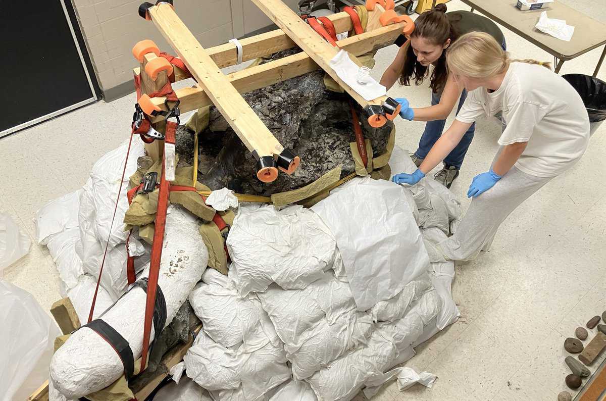
 Non-Invasive Light Spectroscopy Technique Tells a lot about Mechanical Properties of Pharmaceutical Materials, Tissues, Cells and Many More

Sanika Jadhav
College of Pharmacy
Pharmaceutical Sciences and Experimental Therapeutics
Category: Graduate Student
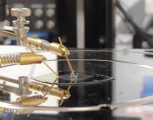
The image is taken while performing Brillouin Light Scattering (BLS) spectroscopy analysis on pharmaceutical materials. This light spectroscopy technique uses a laser as a light source, and multiple mirrors to reflect light and reduce the intensity to appropriate energy before hitting on sample. After hitting the sample the light scatters and it is detected by the detector to interpret the change in energy of scattered light to understand microscopic and macroscopic properties of materials. This spectroscopy relies on the interaction between the light intensity from the laser and sound energy present in the sample to provide relevant information regarding the sample. This technique has huge potential to understand the mechanical properties of a wide variety of pharmaceutical materials ranging from drugs, tissues, cells, and even stiff materials such as animal bones. This is a non-destructive, quick, convenient method and requires a sample in milligrams to run the analysis. Also, once the spectra are recorded for the sample you can use the same spectra to compare it against multiple samples measured at different time points. The Brillouin Light Scattering (BLS) spectroscopic instrument located in Dr. Lewis Stevens’s lab at the College of Pharmacy is one of the very few (likely less than 10) instruments available across the United States. This state-of-the-art instrument has great potential to investigate the mechanical stiffness of pharmaceutical materials and correlate with various intrinsic and macroscopic properties.
Acknowledgments: I would like to thank Dr. Lewis Stevens for setting up this instrument and teaching me how to run it.
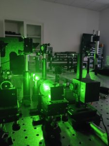
 Engineered Novel Multifunctional Cocrystal of Permeation Enhancer with Pharmaceutical Properties
Sanika Jadhav
College of Pharmacy
Pharmaceutical Sciences and Experimental Therapeutics
Category: Graduate Student
The image was taken using a scanning electron microscope (SEM) located at the Materials Analysis, Testing, and Fabrication (MATFab) facility at the University of Iowa. The image is of novel and unique pharmaceutical material complex with improved attributes we generated in our lab. This material would be game-changing in improving the delivery of pharmaceutical peptides and proteins using the oral route instead of injections. Permeation enhancers are used to improve physiological stability and permeability across cell membranes of various peptides and proteins. However, the powder form of these permeation enhancers is very difficult to handle while making oral tablet dosage forms at the manufacturing scale due to various intrinsic issues such as poor flow. We have generated this unique and multifunctional molecular complex of permeation enhancer by combining it successfully with pharmaceutically safe ingredient. This novel material exhibits significantly improved properties such as very good powder flow and strong, robust tablets. This image shows that the novel pharmaceutical complex has changed its powder morphology and shape compared to the starting materials. This change in powder particle morphology resulted in improved pharmaceutical properties of this new complex. Overall, particle engineering was successfully accomplished by changing the solid forms of an ingredient to improve its ability to deliver oral peptides.
 Modeling Radiologists’ Visual Search in Breast Cancer Screening
Karthika Kelat
College of Engineering
Biomedical Engineering & Radiology – Medical Image Perception Laboratory
Category: Graduate Student
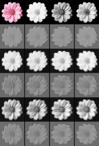
Human perception plays a crucial role in clinical decision-making when interpreting medical images. This image depicts a simulation of how the human visual system processes information. This is fundamental to human perception, enabling us to recognize shapes, textures, and patterns in complex scenes. I used a pink flower here because my research focused in the area of breast cancer. Breast cancer is the most common type of cancer in women. In general,10% to 30% of breast cancers per year are missed at screening. We want to help radiologists commit fewer diagnostic errors by modeling their visual search and the areas where they indicate a lesion is present using what is known about human visual system and machine learning. My research aims to advance the understanding of radiologists’ visual perception in 2D digital mammograms and 3D digital breast tomosynthesis.
Acknowledgments: Professor Claudia Mello-Thoms
 Foaming Synoviovytes
Lauren McNally
College of Liberal Arts and Sciences
Biology – Orthopedics and Rehabilitation
Category: Undergraduate Student
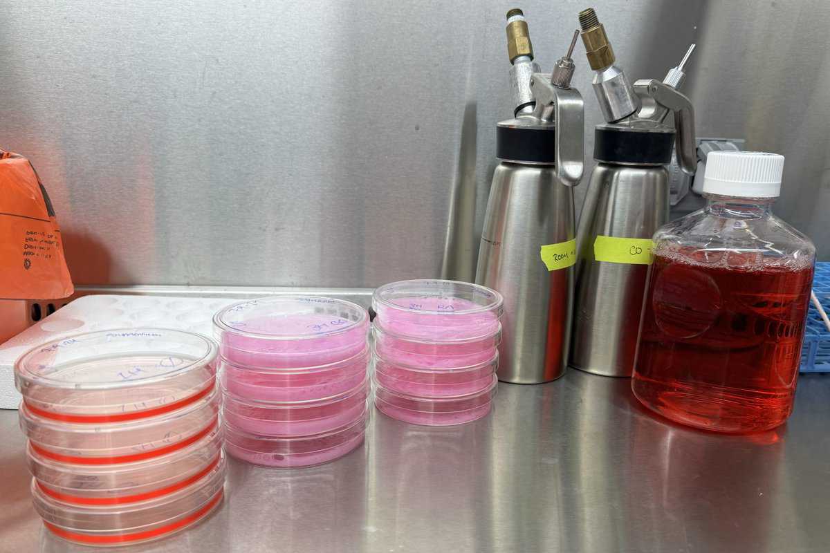
In the image, I worked with synoviocytes (synovium cells) from a bovine model and plated these cells on twelve dishes in a monolayer. I induced oxidative stress with tert-butyl hydroperoxide, mimicking the stress cells experience with injury. The cells’ injury response involves lipid peroxidation, which results in oxidative stress. I am studying the pro-healing effects of carbon monoxide foam on the injury response and mitochondrial activity in synoviocytes. During this experiment, four plates of cells were foamed with carbon monoxide foam (middle stack), four foamed with room air foam (right), and four had a media change (left). The media change acts as a negative control because no foam is applied, so the injury response should occur as normal and provide a baseline. The room air foam harms the injury response, so the cells should show increased signs of stress compared to the negative control. The carbon monoxide is our treatment group of interest, and we have observed increased mitochondrial activity, which promotes cell recovery. On the right of the image, you can see the two foam canisters and cell media solution that provides nutrients for the cell cultures. This image summarizes my research as most of my studies investigate the impact of foam treatments on monolayers of cells (chondrocytes or synoviocytes). After foaming, I stain and image the live cells using confocal microscopy.
Acknowledgments: Dr. Mitchell Coleman
 Hawkeye Pride in Yeast Research
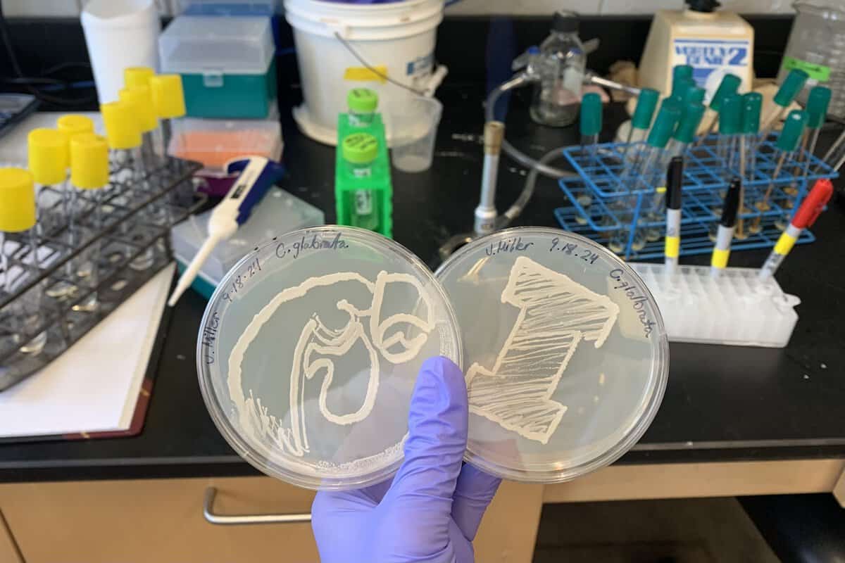
Jessica Miller
College of Liberal Arts and Sciences
Biology – Gene Regulatory Evolution Lab, Dr. Bin He
Category: Undergraduate Student
C. glabrata is recognized as the leading cause of mortality due to fungal infections worldwide. This underscores its critical importance in the field of biological research, particularly in the study of stress responses. The image depicts the species streaked on synthetic complete media, which was cultivated for two days at room temperature. The intricate designs were meticulously applied over a flame, showcasing not only technical skills but also the creativity and dedication that research demands. The level of detail and precision is emblematic of the commitment seen at the undergraduate level, where students embody the Hawkeye spirit of growth and innovation.
Acknowledgments: I want to extend my heartfelt thanks to the world’s best lab members for their unwavering encouragement and positivity!
 Vertical Registration of Atmospheric Layers such as Clouds and Aerosols
Bolaji Oladipo
College of Engineering
Chemical and Biochemical Engineering – Iowa Atmospheric Development Laboratory
Category: Graduate Student
The atmosphere, weather, and climate conditions are changing, and models are failing to accurately predict or escalate extreme weather events such as floods and tornadoes. Even when models do make predictions, they are often far from reality. This was evident in the recent hurricane and flooding in North Carolina. Additionally, in 2022 alone, 3.4 million Americans were displaced from their homes due to extreme weather events, and the U.S. government has spent 1 trillion dollars on damages over the past seven years. The issue lies with the data used for predictions. There is a need for data that can accurately identify both visible and invisible things in the atmosphere and label them correctly—that’s where I come in. We fly lidar-based sensor instruments into the sky, capturing images much like taking pictures with your phone camera. The raw images are gathered and processed to distinguish between visible things like clouds and invisible particles like aerosols. The current method struggles with removing noise that obscures the raw images, which is the light from the sun and excessive noise removal reduces the ability of the images to show small details. What I am doing differently is processing the images in their raw form by training an AI, which works like human eyes. Just as your eyes can distinguish between cats and dogs, my AI was trained to distinguish between clouds and aerosols. The overall goal is to provide data for scientists to feed into their models, enabling them to make accurate and timely predictions of extreme weather events such as floods and tornadoes, and eventually produce real-time data in the future. Knowing what the weather will do tomorrow can help us prepare today—and I hope that one day, we’ll be able to predict the reality of extreme weather with precision.
Acknowledgments: Dr. Joe Gomes and Dr. Matthew McGill
 Flux at Sunrise
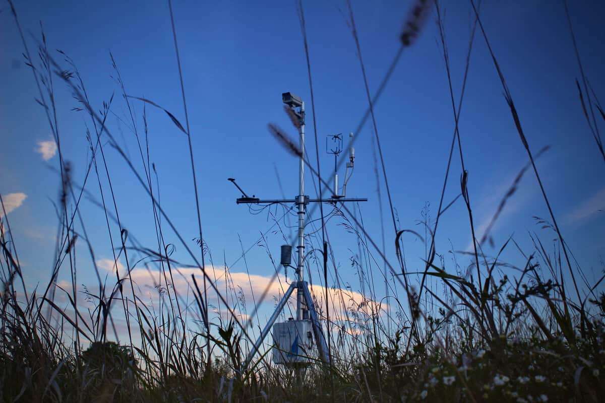
Riley Pacer
College of Liberal Arts and Sciences
Geographical & Sustainability Sciences – ECH2O & LOQATE Labs
Category: Graduate Student
Tallgrass prairies once covered more than 80% of Iowa’s landscape. Today, less than 0.1% of these prairies remain. In 2018, UI students, faculty, and staff began working to restore a portion of Ashton Cross Country Course to the active and biodiverse prairie we enjoy today. Iowa’s remnant and restored prairies provide many critical ecosystem services. They improve soil structure, support nutrient and carbon cycling, and are home to hundreds of plant, animal, and insect species. Additionally, prairies offer local cooling potential, reducing surface and air temperatures through evaporative cooling. My photograph shows the eddy covariance flux tower located at Ashton Prairie Living Laboratory here on UI’s campus. The flux tower continuously measures temperature, carbon, and water fluctuations, enabling us to understand how restored prairies are responding to climate variability. The data collected here will also shed light on the role that prairies play in reducing local temperatures. Through my research, I hope to understand how prairie restoration can play a part in mitigating the impacts of anthropogenic warming.
Acknowledgments: Matt Dannenberg, Susan Meerdink, Nicholas Simons
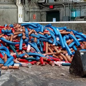 Food Waste to Fuel
Maclaine Putney
College of Engineering
IIHR—Hydroscience & Engineering – IWWERP – IWWERP
Category: Faculty/Staff/Researchers
Ever wonder what happens when food doesn’t sell? Or when grocery stores have an issue that results in total shipments of food spoiling and being unfit for human consumption. Well, the normal solution is the landfill, all that food waste gets dumped in a hole and covered with dirt where it will rot away and produce climate changing gasses like methane and carbon dioxide. The research being conducted in the Iowa Wastewater and Waste to Energy Program aims to help transform those once wasted calories into positive green energy. The facility picture is called The MORC (or the Muscatine Organic Recycling Center). It is a facility in Muscatine Iowa that de-packages all that once wasted food and puts the resulting slurry into anaerobic digesters to produce biomethane. Active research by out lab is helping to optimize operations of the digesters so that more food waste can become energy instead of trash.
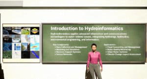 Immersive Learning in Hydroinformatics: AI and Virtual Classrooms for Environmental Education

Ali Rahmani Qeranqayeh
College of Liberal Arts and Sciences
Computer Science – Hydroinformatics Lab
Category: Graduate Student
Our project introduces a groundbreaking virtual classroom that integrates generative AI and immersive technology to enhance Hydroinformatics education. Using a lifelike digital avatar instructor, the system offers interactive and adaptive learning experiences where students explore complex topics like flood forecasting, water quality monitoring, and climate change assessment in an engaging, personalized way. This immersive environment allows users to interact with real-world hydrology simulations, breaking traditional educational barriers. Generative AI tailors the learning process to individual needs, making science education more accessible and effective. As immersive learning technologies become more widespread, this platform is set to democratize education, enabling learners worldwide to access advanced knowledge and better understand critical environmental challenges.
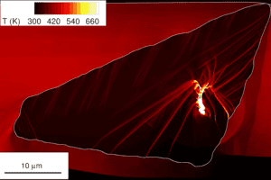 When crystals are in shock!

Shobhan Roy
College of Engineering
Mechanical Engineering – IIHR – Hydroscience & Engineering
Category: Graduate Student
The image shows a simulated snapshot of a single HMX crystal under shock loading. HMX belongs to a class of materials called energetic materials which are used in propellants, explosives, and pyrotechnics. These undergo a shock-to-detonation transition (SDT) when subjected to a sudden strong pressure pulse. This process begins with the formation of high-temperature regions called hotspots, which can grow and propagate into a detonation wave. Our in-house continuum mechanics code, SCIMITAR3D, captures this initial phase of hotspot development in a shocked HMX crystal. SCIMITAR3D is an Eulerian hydrocode that simulates the evolution of multi-material, highly compressive flows over time, incorporating complex physical phenomena such as material interactions, chemical decomposition, phase changes, and elastic and plastic deformations. In this simulation, an HMX crystal embedded in a polymeric binder matrix is impacted by a thin aluminum disc traveling at 1 km/s. The resulting shock wave propagates through the composite material and is visible in the image at 9.2 nanoseconds after the impact as the red-black interface at the bottom of the image. Notable features in this image include sharp needle-like hotspots, known as shear bands, and a bright red hotspot formed by the collapse of a pore in the crystal. Visualizing these features requires high-resolution calculations and accurate numerical models. This research enhances our understanding of the shock initiation process in energetic materials, providing crucial insights for predicting and controlling SDT in practical applications.
Acknowledgments: Jacob Herrin, Prof. H. S. Udaykumar
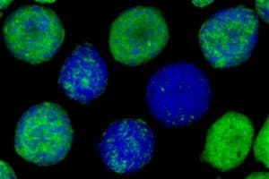 Sheet of “Chocolate Chip Cookies”

Austin Scholp
College of Engineering
Biomedical Engineering – Multi-scale Mechanics, Mechanobiology, and Tissue Engineering Lab
Category: Graduate Student
Wound healing is a complex process that involves the coordination of several different cell types including, fibroblasts, macrophages, and epithelial cells. Adipose tissues also appear to play an important role in wound healing and scar formation. Recent studies have shown that adipocyte lineage cells can accelerate healing and promote extracellular matrix remodeling. We are interested in understanding how the various cell types involved in wound healing interact to produce normal healing. To do this, we have been implementing scaffold-free spherical microtissues, also known as spheroids, to model the interactions between cell types that can occur during the healing process. Two cell types of interest are fibroblasts and pre-adipocytes. We have started by characterizing how these two cell types organize themselves into spheroids. In this image, we see several spheroids made from different ratios of pre-adipocytes and fibroblasts. Fibroblasts are stained green, while pre-adipocytes are stained blue.
 Dentate Gyrus of a Mouse Model of Autism
Stefan Strack
CCOM
Neuroscience & Pharmacology
Category: Faculty/Staff/Researchers
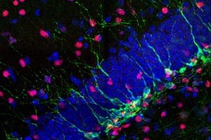
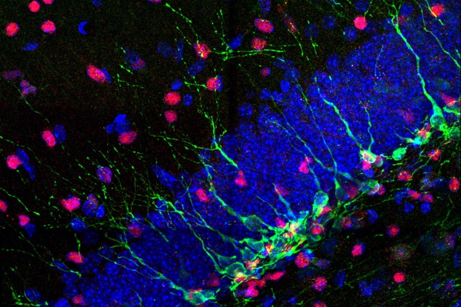
This confocal microscopy image shows a region of the brain (dentate gyrus) of a mouse model of autism labeled with antibodies to two different proteins (red/purple = SOX2; green = DCX) and a blue stain that marks DNA. Different from a normal mouse are the lower number of SOX2-positive nuclei and the more extensive arborization of DCX-positive dendritic trees of neurons. Both SOX2 and DCX are proteins involved in embryonic and adult neurogenesis. The image and its quantification therefore indicate that autism can arise from derailed neurogenesis.
Acknowledgments: Chunling Chen, Chian Ju Jong
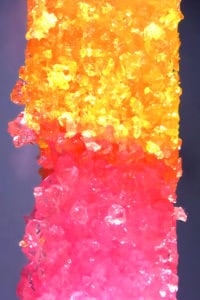 Sweet Interface: Different Sugar Crystal Sizes Bonded Through Polymer to Study the Mechanical Behavior of Dissimilar Interfaces
Nazanin Tabatabaei
College of Engineering
Industrial Engineering – Additive Manufacturing-Integrated Product Realization Laboratory
Category: Graduate Student
This research explores the mechanical behavior of powder-based composites using a simulant polymer-bonded explosives composite comprised of sugar particles of varying sizes. These particles are distinctly dyed pink and yellow to differentiate their granularity, with coarse particles colored pink and fine particles colored yellow. The interface zone between these particles, which emerges during the curing process of the silicon-based polymer, manifests as a distinctive orange hue. The study specifically aims to investigate the effects of this dissimilar interface on the mechanical properties of the composite, providing valuable insights into the interactions within heterogeneous materials.
 Biologically Active Beads to Improve Stormwater Quality
Debojit Tanmoy
College of Engineering
Civil and Environmental Engineering – IIHR – Hydroscience & Engineering
Category: Graduate Student
Green Stormwater Infrastructure (GSI) is becoming popular to manage urban stormwater runoff and improve groundwater infiltration. Even though GSI can efficiently remove particle-bound contaminants, polar trace organic contaminants (TOrCs) and dissolved nutrients can pass through the GSI system and risk groundwater contamination. We developed a novel biologically active engineered geomedia (BioSorp Bead) that could be used in existing and/or new GSI for rapid sorption and subsequent biodegradation of stormwater-relevant contaminants, including TOrCs and dissolved nutrients. The beads could be used to bioaugment GSI to bioremediate the sorbed pollutants in situ. Thus, biodegradation could renew GSI sorption capacities, and increase the service-life sustainability. The BioSorp Bead is a cation-alginate composite where we encapsulate black carbon, mineral sorbents, and microorganisms for contaminant biodegradation (white rot fungi as model organisms). We also add a supplemental carbon source and external electron shuttles to aid fungal viability, activity, and degradation. The beads are made of non-toxic, economical recycled materials and the production process is easily scalable for field application.
Photograph: White-rot fungi growing from the beads
Acknowledgments: Gregory H. LeFevre
 Record Player for Science
Fatima Toor
College of Engineering
Electrical and Computer Engineering – Iowa Technology Institute
Category: Faculty/Staff/Researchers
The photo shows an electrical probe station in our lab that allows us to measure electrical properties of sensors and other devices made in our lab. The shot is taken during a measurement of the sensor response with an analyte drop present on the nanostructured sensor area. The sensor is made of silicon, a common material for microelectronics, and being used to measure the concentration of estrogen in a buffer solution. At the time this photo was taken, we were working with Prof. Greg LeFevre in Environmental Engineering looking to develop sensors for detecting estrogen levels in waterways. From afar, the image looks like a record player, playing some science rhythms.
Acknowledgments: Wenqi Duan, a past graduate student, who supported the biosensor development work
 At the Reservoir’s Edge
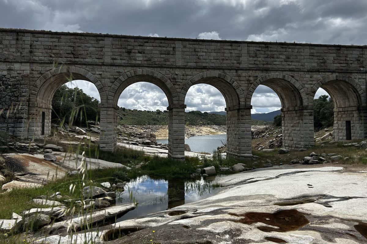
Corinne Watts
College of Liberal Arts
Anthropology
Category: Graduate Student
The photo shows an arched support as part of the Embalse del Atazar, a critical water reservoir in Comunidad de Madrid, Spain. This reservoir, located near the rocky Sierra Norte, was built between 1968 and 1972 to prevent the depletion of water reserves for Madrid, including the populous metro area. I captured this photo while collecting rock samples for my geoarchaeological dissertation research. My project examines how prehistoric (Neolithic and Chalcolithic) communities collected raw materials to create stone tools, objects critical to survival, economic connection, and identity. This project brought me to the edge of several bodies of water, including this reservoir, as they are critical for communities living in the hot and dry landscape in this area of Spain, just as they were in the past. Water is vital to all human activities and was especially important in this region as communities began to rely on domesticated plants and animals in prehistory. When I took this photo, it was near the middle of summer and the reservoir was not fully depleted as recent rains had bolstered the water levels. These levels fluctuate throughout the year and have been impacted by the rising temperatures and increasing aridity in Spain. Bodies of water are integral to human survival and are deeply connected to our experiences. Critically, with the landscape captured in this photo, I see a connection between the past, which I study, and the present, where I conduct research.